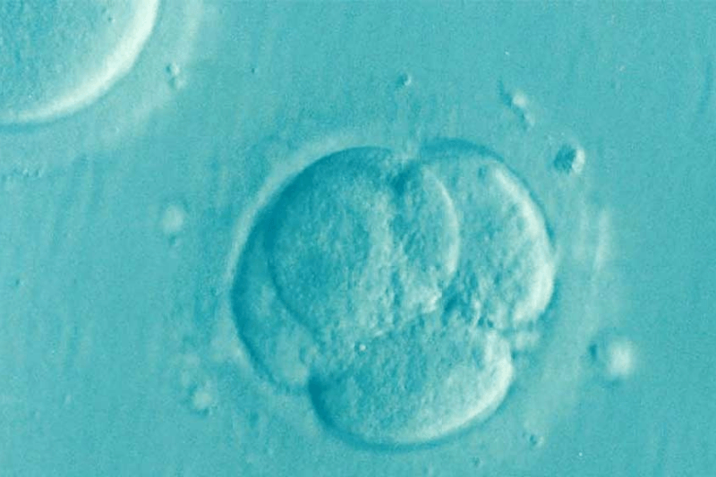
What is In Vitro Fertilization (IVF)?
In vitro fertilization (IVF) is defined as any fertility treatment where both an egg and sperm are removed from the body and used to create an embryo in a lab setting. The embryo is then placed back into the uterus to grow and develop, just like in a natural pregnancy. For anyone struggling to start a family, IVF has represented new hope since it was first introduced nearly 40 years ago.
While women whose IVF treatments have resulted in multiple births seem to get the most media attention, the goal of IVF is to enable couples to have ONE healthy baby at a time, and to potentially create additional embryos that can be frozen for future use.
Who Can Benefit?
Good candidates for IVF may include:
- Women who don’t ovulate on their own, or don’t respond to medications to stimulate ovulation
- Men with sperm abnormalities, such low motility (movement), low sperm count or irregular shape
- Women who have had a tubal ligation or had their fallopian tubes removed
- Men who have had a vasectomy
- Couples whose infertility is still unexplained after extensive testing
- Couples who have exhausted other treatment options without success
- Couples who wish to have genetic testing conducted on their embryo or to screen for future health problems
Success Rates
Success for IVF depends on many different factors, such as the number and quality of the eggs and sperm. Success rates also decrease as women age, but IVF has the highest success rate of all the fertility treatments.
Overall, in women under age 35, the success for achieving a pregnancy with one IVF cycle is about 50%. The live birth rate for IVF is lower because of the risk for miscarriage or ectopic pregnancy.
Preimplantation Genetic Testing (PGT)
Embryos produced by IVF are studied using a special microscope to predict their ability to produce a pregnancy by their overall appearance. However, this method does not reliably predict whether or not an embryo will result in a successful pregnancy.
Preimplantation genetic testing, or PGT, is a technique in which a small sample of cells are removed from the embryo, and these cells are then used to evaluate the genetic composition of the embryo as a whole. Cells removed from each developing embryo are used for genetic analysis to determine if the embryo is affected by chromosomal defects which would impact implantation, miscarriage, and may cause subsequent birth defects.
How is the Biopsy Performed for PGT?
In order to detect genetic defects, a small sample of the embryo must be biopsied and sent to an independent genetics lab. This sample of cells contains chromosomes, and the number of chromosomes will be analyzed by the genetics lab. In order to be eligible for biopsy, embryos must have continued to develop 5 and 6 days after your retrieval. After a small group of cells is removed from the blastocyst, the embryo is frozen. This halts the embryo’s development indefinitely, and allows time for the genetics lab to receive the cell sample and screen for abnormalities. The screening process can text 2-3 weeks.
At the genetics lab, technologists compare the sample from each embryo to a standard that is known to be normal. When finished, a report is sent to our lab indicating which embryos have the correct number of chromosomes (also known as “euploid”). As part of this testing, the 23rd pair of chromosomes is analyzed. This pair determines the sex of the embryo, so the report can indicate if the embryo will develop into a male or female, if this information is desired.
Once we have received the genetics report, we will know which of the cryopreserved embryos are genetically normal. A genetically normal embryo can then be thawed and transferred as part of a frozen embryo transfer cycle. Pregnancy rates are generally higher when transferring embryos that have been screened via PGT as the embryo for transfer does not have any chromosomal defects.
What is the difference between PGT-A and PGT-M?
PGT-A is a test that looks for large genetic abnormalities. The “A” stands for aneuploidy, a term used to denote an abnormal number of chromosomes. These abnormalities are common in embryos, and their occurrence increases as the biological mother ages. PGT-A can only detect when large pieces of chromosomes or entire chromosomes are missing or duplicated.
PGT-M is a more detailed test that looks for a very specific genetic abnormality. The “M” stands for monogenic disorders, which are caused by variation in a single gene. Examples of monogenic disorders include sickle cell disease, Huntington’s disease, and cystic fibrosis. In order to perform this test, the genetics lab needs to know which disease they are looking for ahead of time. This is generally indicated by a patient’s carrier screening tests or if they have a family member who expresses a disease caused by a specific genetic defect. When the genetic abnormality is known, the genetic material of the embryo can be analyzed to see if that embryo could result in the birth of a child with that specific disease. It is not feasible to screen every embryo for every disease-causing mutation; thus, PGT-M is only performed when it is highly likely to improve a patient’s chances of having a healthy baby without a specific disease.
Does Insurance Cover IVF?
We accept most major commercial insurance plans. Feel free to call 502-743-4723 with any questions.
Find out if it’s right for you
While IVF may be the best-known method to treat infertility, most couples don’t need it. Many infertility issues can be corrected with medication or minimally invasive surgery.
If you’re having trouble getting pregnant on your own, contact us to find out if you are a good fit for IVF. We’ll do a complete evaluation to determine if it’s the right choice for you. Contact us today!


